Understanding Baker’s Cyst (Popliteal Cyst)
A Baker’s cyst (popliteal cyst) is a fluid-filled sac that forms behind the knee.
Parts of the knee
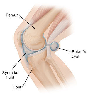
The knee is a complex joint that has many parts. The lower end of the thighbone (femur) rotates on the upper end of the shinbone (tibia). There are several small bursae around the knee joint. These are small sacs filled with a special fluid (synovial fluid) that cushions the rest of the joint. Between the bones is a space that also contains this fluid.
What causes a Baker’s cyst?
It is caused when extra fluid from the knee joint flows into the small bursa that sits behind the knee. When this sac fills with too much fluid, it’s called a Baker’s cyst. This might happen when an injury or disease irritates the knee joint.

In adults, other problems with the knee joint often cause the Baker’s cyst. Injury or a knee disorder such as arthritis can change the normal structure of the knee joint. This can cause a cyst to form.
The synovial fluid inside the joint space may build up as a result of injury or disease. As the pressure builds up, the fluid may bulge into the back of the knee. This can cause the cyst.
Symptoms of a Baker’s cyst
A Baker’s cyst often doesn’t cause symptoms. A cyst will more often be seen on an imaging test, like MRI, done for other reasons. If you do have symptoms, they may include:
-
Pain in the back of the knee.
-
Knee stiffness.
-
Sense of swelling or fullness behind the knee, especially when you straighten your leg.
-
A swelling behind the knee that goes away when you bend your knee.
These symptoms tend to get worse when standing for a long time, or being active.
Diagnosing a Baker’s cyst
Your health care provider will ask you about your medical history and your symptoms. They will give you a physical exam, which will include a careful exam of your knee. It’s important to make sure your symptoms are caused by a Baker’s cyst and not a tumor or a blood clot.
If the cause of your symptoms is not clear, you may have imaging tests, such as:
-
Ultrasound, to look at the cyst in more detail or to look for a blood clot.
-
X-ray, to get more information about the bones of the joint.
-
MRI, if the diagnosis is still unclear after ultrasound, or if your provider is considering surgery.